Send Message

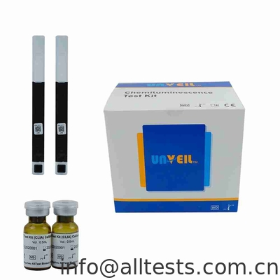
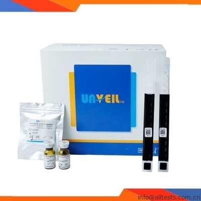
Pepsinogen Ⅱ (PG Ⅱ) Test Kit CLIA Double Antibody Sandwich Method

Basic Information
Place of Origin: China
Brand Name: Unveil
Certification: CE
Model Number: CI-PG Ⅱ
Minimum Order Quantity: N/A
Price: negotiation
Packaging Details: 40T
Delivery Time: 2-4 Weeks
Supply Ability: 10 M tests/month
Product Name: Pepsinogen Ⅱ (PG Ⅱ) Test Kit (CLIA),Double Antibody Sandwich Method,CI-PG Ⅱ Cat No.: CI-PG Ⅱ
Principle: Double Antibody Sandwich Method Specimen: S/P
Pack: 40T Shelf Life: 2 Years
Format: Strip Certificate: CE
Storage Temperature: 2-8℃ Cut-Off: 1.4-200 Ng/mL
High Light:

Pepsinogen Ⅱ CLIA Test

,

PG Ⅱ Chemiluminescence Immunoassay Test

,

CLIA Test Double Antibody Sandwich Method

Pepsinogen Ⅱ (PG Ⅱ) Test Kit (CLIA),Double Antibody Sandwich Method,CI-PG Ⅱ
 
A CLIA test kit for quantitative determination of PepsinogenⅡ (PGⅡ) in human serum or plasma with the use of Automatic Chemiluminescence Immunoassay Analyzer.
 
Product Name: Pepsinogen Ⅱ (PG Ⅱ) Test Kit (CLIA),Double Antibody Sandwich Method,CI-PG Ⅱ
Principle: Double Antibody Sandwich Method
Pack: 40T
Format: Strip
Storage Temperature: 2-8℃
Cat No.: CI-PG Ⅱ
Specimen: S/P
Shelf Life: 2 Years
Certificate: CE
Cut-Off: 1.4-200 Ng/mL


【INTENDED USE】
 
The PepsinogenⅡ (PGⅡ) Test Kit (CLIA) is intended for quantitative determination of PepsinogenⅡ (PGⅡ) in human serum and plasma, as an aid in diagnosis of stomach disease in clinical practice.
For professional in vitro diagnostic use only.
 
【SUMMARY】
 
Pepsinogen (PG) is the precursor of pepsin, which is a digestive enzyme specifically produced in the gastric mucosa. The human stomach expresses two isoenzymes, PG Ⅰ and PG Ⅱ , which have different biochemical and immune properties. Histological studies based on immunohistochemistry using specific antibodies or in situ hybridization clearly identified PGI and PGII producing cells. PGⅠis mainly secreted by the principal cells of the fundus glands and mucous neck cells, but PGⅡ is produced not only in these cells, but also in the Brunner gland cells of the heart, pylorus and duodenum.
 
Pepsinogen is mainly excreted into the gastric cavity, but about 1% of it enters the bloodstream. Studies have clarified that the serum PG level reflects the morphology and function of the gastric mucosa and various pathological conditions, such as inflammation. It is worth noting that in the process of chronic atrophic gastritis, mucosal atrophy progresses from the pyloric gland side to the oral side, and the level of PGⅠ and the ratio of PGⅠ /PG Ⅱ decrease with the progress of mucosal atrophy.
 
These clinically important changes in serum PG levels are due to the unique distribution of the above-mentioned PG-producing cells in the epithelial cells of the gastric mucosa. In addition, the results of past pathological and epidemiological studies have shown that there is a strong correlation between chronic atrophic gastritis and differentiated gastric cancer.
 
Therefore, chronic atrophic gastritis is considered to be a precancerous lesion. The pepsinogen test is based on the correlation between chronic atrophic gastritis and the development of gastric cancer on the one hand and on the other hand is more based on the correlation between chronic atrophic gastritis and low PG levels.
 
By introducing a PG test that can identify subjects at high risk for gastric cancer, and by introducing an endoscopy for screening subjects with a positive PG test, the efficiency of gastric cancer detection can be significantly improved.
 
【PRINCIPLE】
 
This product uses the double antibody sandwich method. In the first step, the sample, the PGⅡ antibody labeled with alkaline phosphatase, and the magnetic particles coated with the PG Ⅱ antibody are mixed. After incubation, the PGⅡ in the sample forms an immune complex with the corresponding antibody. In the second step, magnetic separation and cleaning are performed to remove free enzyme-labeled antibodies. The third step is to add the chemiluminescent substrate solution to the immune complex. Luminescence signal generated by the enzyme reaction is detected by Automatic Chemiluminescence Immunoassay Analyzer and the detected luminescence intensity is related to the concentration of PGⅡ in the sample. Automatic Chemiluminescence Immunoassay Analyzer can calculate the concentration of PGⅡin the sample.
 
【REAGENTS】
 
The reagent strip includes PGⅡ antibody coated with magnetic particles, alkaline phosphatase labeled PGⅡ antibody, wash buffer and substrate solution.
 
【STORAGE AND STABILITY】
 
1. Unopened test kits should be stored at 2-8 °C. When stored and handled as required, all unopened reagents are stable through the expiration date printed on the label.
 
2. Do not freeze. Do not turn the reagent strips over.
 
3. Store the test kit upright. Do not expose reagents to strong light during storage. Care should be taken to protect the components of the test from contamination.
 
4. Remaining reagents in the kit should be stored at 2-8°C immediately.
 
5. Do not use if there is evidence of microbial contamination or precipitation. Biological contamination of dispensing equipment,
containers or reagents may lead to false results.
 
6. Calibrator and Control Materials: Unopened:
Stable until the expiration date when stored at 2-8 °C.
Opened:
Stable for 1 week at 2-8 °C
 
【SPECIMEN COLLECTION AND PREPARATION】
 
Collect and handle the specimen strictly as below:
 
1. Collect the specimen according to standard procedures.
 
2. Human serum collected using standard sampling tubes or tubes containing separating gel.
 
3. Human plasma collected using EDTA or Heparin sodium.
 
4. Separate human serum and plasma from blood as soon as possible to avoid hemolysis. Grossly hemolytic, lipidic or turbid samples should not be used. Specimen with extensive particulate should be clarified by centrifugation prior to use. Do not use specimens with fibrin particles or contaminated with microbial growth.
 
5. Do not leave specimens at room temperature for prolonged periods. Serum and Plasma specimens may be stored at 2-8 °C for up to 5 days, below -20 °C stable for 3 months.
 
6. Bring specimens to room temperature prior to testing. Frozen specimens must be completely thawed and mixed well prior to testing.Specimens should not be frozen and thawed repeatedly.
 
7. If the patients had been treated by high dose of biotin (5 mg/day), blood collection must be done after at least 8 hours.
 
8. If specimens are to be shipped, pack them in compliance with local regulations covering the transportation of etiologic agents.

Contact Details
selina

Phone Number : +8615857153722

WhatsApp : +8613989889852