Send Message

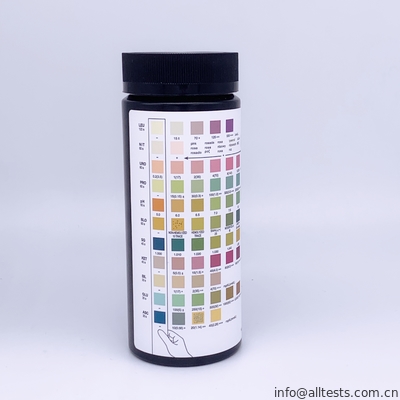
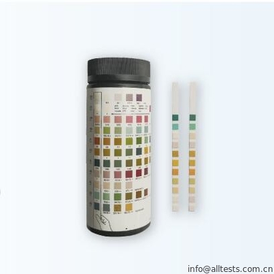
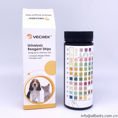
Veterinary Urinalysis Strips With Fast Reading For rapid detection of multiple analytes in animal urine.

Basic Information
Place of Origin: CHINA
Brand Name: AllTest
Certification: CE
Model Number: U031-01-14
Minimum Order Quantity: 500
Packaging Details: 40T/kit
Supply Ability: 100 Million a year
Format: Strip Specimen: Urine
Kit Size: 100T/kit Cut-Off: See Insert
Storage: 2-30℃ Shelf Time: 24 Months
High Light:

rapid diagnostic test kits

,

One Step Rapid Test

For rapid detection of multiple analytes in animal urine. For in vitro diagnostic use only.


Application and description:


The Urinalysis Reagent Strips (Urine) are firm plastic strips onto which several separate reagent areas are affixed. The test is for the qualitative and semi-quantitative detection of one or more of the following analytes in urine: Ascorbic acid,Glucose, Bilirubin,Ketone (Acetoacetic
acid), Specific Gravity, Blood, pH, Protein, Urobilinogen, Nitrite and Leukocytes. The Urinalysis Reagent Strips (Urine) are for single use in professional near-patient (point-of-care) and centralized laboratory locations.
Refer to kit box label for the specific analyte(s) listed, and compare to the appropriate analyte(s) and color blocks on the color chart for results.


Description:


Ascorbic acid: This test involves decolorization of Tillmann’s reagent. The presence of ascorbic acid causes the color of the test field to change from blue-green to orange. Patients with adequate diet may excrete 2-10 mg/dL daily. After ingesting large amounts of ascorbic acid,
levels can be around 200 mg/dL.
Glucose: This test is based on the enzymatic reaction that occurs between glucose oxidase, peroxidase and chromogen. Glucose is first oxidized to produce gluconic acid and hydrogen peroxide in the presence of glucose oxidase. The hydrogen peroxide reacts with potassium
iodide chromogen in the presence of peroxidase. The extent to which the chromogen is oxidized determines the color which is produced, ranging from green to brown. Glucose should not be detected in normal urine. Small amounts of glucose may be excreted by the kidney.
Glucose concentrations as low as 100 mg/Dl may be considered abnormal if results are consistent.
Bilirubin: This test is based on azo-coupling reaction of bilirubin with diazotized dichloroaniline in a strongly acidic medium. Varying bilirubin levels will produce a pinkish-tan color proportional to its concentration in urine. In normal urine, no bilirubin is detectable by even the most
sensitive methods. Even trace amounts of bilirubin require further investigation. Atypical results (colors different from the negative or positive color blocks shown on the color chart) may indicate that bilirubin-derived bile pigments are present in the urine specimen, and are possibly
masking the bilirubin reaction.
Ketone: This test is based on ketones reacting with nitroprusside and acetoacetic acid to produce a color change ranging from light pink for negative results to a darker pink or purple color for positive results. Ketones are normally not present in urine. Detectable ketone levels
may occur in urine during physiological stress conditions such as fasting, pregnancy and frequent strenuous exercise. In starvation diets, or in other abnormal carbohydrate metabolism situations, ketones appear in the urine in excessively high concentration before serum ketones
are elevated.
Specific Gravity: This test is based on the apparent pKa change of certain pretreated polyelectrolytes in relation to ionic concentration. In the presence of an indicator, colors range from deep blue-green in urine of low ionic concentration to green and yellow-green in urine of
increasing ionic concentration.
Blood: This test is based on the peroxidase-like activity of hemoglobin which catalyzes the reaction of diisopropylbenzene dihydroperoxide and 3,3',5,5'-tetramethylbenzidine. The resulting color ranges from orange to green to dark blue.
pH: This test is based on a double indicator system which gives a broad range of colors covering the entire urinary pH range. Colors range from orange to yellow and green to blue.
Protein: This reaction is based on the phenomenon known as the “protein error” of pH indicators where an indicator that is highly buffered will change color in the presence of proteins (anions) as the indicator releases hydrogen ions to the protein. At a constant pH, the
development of any green color is due to the presence of protein. Colors range from yellow to yellow-green for negative results and green to green-blue for positive results.
Urobilinogen: This test is based on a modified Ehrlich reaction between p-diethylaminobenzaldehyde and urobilinogen in strongly acidic medium to produce a pink color.
Nitrite: This test depends upon the conversion of nitrate to nitrite by the action of Gram negative bacteria in the urine. In an acidic medium, nitrite in the urine reacts with p-arsanilic acid to form a diazonium compound. The diazonium compound in turn couples with 1
N-(1-naphthyl) ethylenediamine to produce a pink color.
Leukocytes: This test reveals the presence of granulocyte esterases. The esterases cleave a derivatized pyrazole amino acid ester to liberate derivatized hydroxyl pyrazole. This pyrazole then reacts with a diazonium salt to produce a beige-pink to purple color. Normal urine
specimens generally yield negative results. Trace results may be of questionable clinical significance.

 
How to use?
 

DIRECTIONS FOR USE
Allow the strip, urine specimen, and/or controls to reach room temperature (15-30ºC) prior to testing.
1. Remove the strip from the closed canister and use it as soon as possible. Immediately close the canister tightly after removing the required number of strip(s). Completely immerse the reagent areas of the strip in fresh, well-mixed urine and immediately remove the strip to avoid
dissolving the reagents. See illustration 1 below.
2. While removing the strip from the urine, run the edge of the strip against the rim of the urine container to remove excess urine. Hold the strip in a horizontal position and bring the edge of the strip into contact with an absorbent material (e.g. a paper towel) to avoid mixing chemicals from adjacent reagent areas and/or soiling hands with urine. See illustration 2 below.
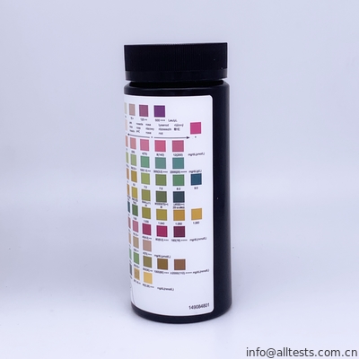
3. Compare the reagent areas to the corresponding color blocks on the canister label at the specified times. Hold the strip close to the color blocks and match carefully. See illustration 3 below.
Note: Results may be read up to 2 minutes after the specified times. Results may also be read using the Urine Analyzers. Refer to the Instruction Manual for details.


 Veterinary Urinalysis Strips With Fast Reading For rapid detection of multiple analytes in animal urine. 0


INTERPRETATION OF RESULTS
Results are obtained by direct comparison of the color blocks printed on the canister label. The color blocks represent nominal values; actual values will vary close to the nominal values. In the event of unexpected or questionable results, the following steps are recommended: confirm that the strips have been tested within the expiration date printed on the canister label, compare results with known positive and negative controls and repeat the test using a new strip. If the problem persists, discontinue using the strip immediately and contact your local distributor.

Contact Details
selina

Phone Number : +8615857153722

WhatsApp : +8613989889852